Harness machine learning and computer vision to detect infected blood cells, accelerate malaria diagnosis, and deliver accurate results to support clinical decision-making.
Client Overview
A network of clinics operating in high-malaria-prevalence regions of sub-Saharan Africa was relying entirely on manual microscopy for malaria diagnosis. Trained laboratory technicians examined Giemsa-stained blood smear slides under a microscope, manually identifying and counting infected red blood cells to diagnose and determine the severity of malaria infection. The process was slow — a thorough examination of a single slide could take 20 to 45 minutes — and throughput was limited by the number of trained technicians available at each clinic site, several of which were serving large catchment populations in remote areas with limited staffing capacity. The quality and consistency of diagnoses varied significantly between technicians and deteriorated with fatigue, particularly during high-volume periods and in clinics where a single technician was responsible for all laboratory work. Malaria is a life-threatening illness where treatment delay measurably worsens outcomes, and in resource-constrained settings where parasite rapid diagnostic tests were not always available or reliable, microscopy remained the gold standard — meaning diagnostic capacity directly constrained treatment capacity. The clinic network's clinical directors wanted to explore whether AI-assisted microscopy analysis could augment their limited technician capacity — not replacing the skilled human review that remained essential for difficult cases, but accelerating the initial screening process and reducing the time technicians spent on routine examination of clearly negative slides or clear-cut positive cases with high parasite density.
Our Approach
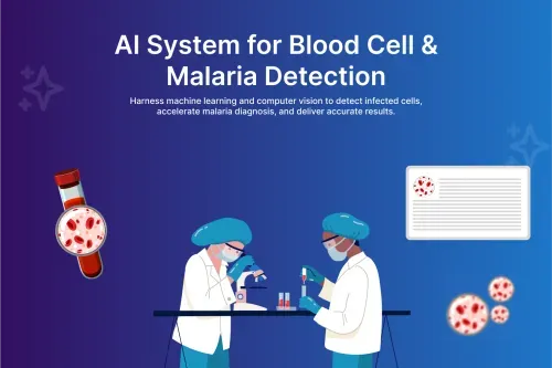
Zentric Solutions developed a convolutional neural network-based malaria detection system using TensorFlow and Keras, trained on a large dataset of annotated blood smear images containing both Plasmodium-infected and healthy red blood cells. The model was designed to detect and classify individual red blood cells within a blood smear image, identifying infected cells with parasites at the trophozoite, schizont, and gametocyte stages, and calculating a parasite density estimate based on the ratio of infected to total cells detected in the examined image area. The inference pipeline was deployed on AWS SageMaker, accessible from the clinic sites via a lightweight web application built in React. Laboratory technicians photographed prepared blood smear slides using a calibrated smartphone camera adapter mounted to their existing microscopes, uploading the images through the web interface for analysis. The AI system returned a cell-level detection result — with infected cells highlighted by bounding box overlays on the original image — alongside a parasite density percentage, a preliminary diagnosis classification (negative, low density, moderate density, or high density), and confidence scores for each classification. The system was explicitly designed to support rather than replace technician judgement. All AI results were presented as preliminary screening outputs to be reviewed and confirmed by the technician before recording a clinical diagnosis. A result review interface allowed technicians to accept the AI classification, override it with their own assessment, or flag the slide for a second opinion. Overrides and flagged cases were collected as a continuous learning dataset to support ongoing model improvement. Processing time per slide through the AI pipeline was under 90 seconds from image upload to result display, dramatically reducing the time technicians spent on routine screening and freeing their attention for cases requiring detailed expert examination.
Tech Stack
Project Tags
Everything you need to know about this project and our approach.
Zentric Solutions delivers cutting-edge digital products that streamline operations, enhance engagement, and drive lasting growth.